How Fluorescent Signals Reveal Cellular Biology
Discover how fluorescence microscopy transforms molecular signals into biological images, quantitative measurements, and insights across modern life science research.
Fluorescence Microscopy:
How Fluorescent Signals Reveal Cellular Biology
Fluorescence microscopy transforms molecular signals into visible patterns, allowing researchers to observe cellular structures, follow biological processes, and convert images into quantitative biological information.
Fluorescence imaging connects optical excitation, molecular emission, detection, and biological interpretation.
What Is Fluorescence Microscopy?
Fluorescence microscopy is an optical imaging approach that uses fluorescent molecules to make specific biological structures or molecular events visible. Instead of relying only on the natural appearance of a cell, researchers can detect fluorescent signals associated with proteins, nucleic acids, membranes, organelles, or other biological targets.
The principle is based on the interaction between light and a fluorophore. A fluorophore absorbs light at particular wavelengths and then releases part of that energy as light at a longer wavelength. This separation between excitation and emission allows the microscope to illuminate the sample while selectively detecting the resulting fluorescent signal.
Fluorescence microscopy converts molecular properties into spatially resolved optical signals that can be observed, recorded, and quantified.
How Fluorescent Signals Are Generated
A fluorescent molecule does not simply produce light continuously. Its electronic structure determines which wavelengths it can absorb and which wavelengths it can emit. When excitation light reaches the fluorophore, electrons are temporarily promoted to a higher-energy state.
As the molecule returns toward its ground state, part of the absorbed energy is released as fluorescence. This creates the characteristic separation between excitation and emission wavelengths.
Excitation
The fluorophore absorbs energy from incident light.
Emission
The molecule releases part of that energy as fluorescent light.
Detection
Optical filters and detectors isolate and record the signal.
Fluorophores and Fluorescent Probes
The biological usefulness of fluorescence depends heavily on the probe used to generate the signal. Fluorophores can be incorporated into antibodies, small-molecule probes, nucleic-acid probes, or genetically encoded proteins. The choice of probe determines which biological information can be observed.
Genetically encoded fluorescent proteins such as GFP and engineered variants have become particularly important because they allow researchers to connect fluorescence directly to protein expression or localization inside living cells.
Figure 1. Fluorescence microscopy connects excitation, optical collection, detection, and molecular visualization. Scientific source →
From Fluorescent Molecules to a Microscope Image
A fluorescence microscope transforms molecular emission into a digital image through a sequence of optical and electronic processes. Excitation light is delivered to the sample, fluorescent emission is collected by the objective, and optical components separate the desired emission from unwanted wavelengths.

Figure 2. Light-sheet fluorescence microscopy uses selective illumination to improve optical sectioning and enable advanced biological imaging. Nature Methods source →
Choosing the Right Imaging Strategy
No single fluorescence microscopy method is optimal for every biological question. Widefield microscopy can provide rapid imaging across large fields, while confocal approaches improve optical sectioning by rejecting out-of-focus light.
Light-sheet approaches selectively illuminate the sample and are particularly useful for three-dimensional and live imaging.

Figure 3. Light-sheet fluorescence imaging demonstrates how selective illumination can be applied to dynamic three-dimensional biological samples. Nature Methods source →
How Microscopy Generates Biological Data
A fluorescence image contains far more information than its visual appearance suggests. Each pixel represents a numerical measurement of intensity, while multiple fluorescence channels can encode different molecular targets.
A time series can additionally reveal changes in localization, morphology, or signal intensity. Modern automated microscopes can therefore generate large datasets rather than isolated pictures.
Image → Data
Intensity, size, shape, localization, spatial relationships, and temporal changes can all become measurable biological variables.
From Images to Quantitative Measurements
Quantitative image analysis transforms fluorescence microscopy from a primarily visual technique into a measurement platform. Researchers can identify cells or subcellular structures, measure fluorescence intensity, calculate object size, characterize morphology, and investigate spatial relationships.
A typical computational workflow can involve illumination correction, segmentation, object identification, feature extraction, and statistical analysis.
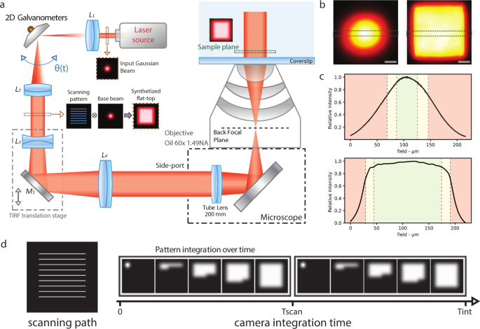
Figure 4. Uniform illumination is important when fluorescence intensity is used as a quantitative measurement across a field of view. Nature Communications source →
Super-Resolution and the Search for Molecular Detail
Conventional fluorescence microscopy is constrained by the diffraction of light. Modern super-resolution approaches overcome or circumvent aspects of this limitation by controlling how fluorescent molecules are excited, detected, or localized.
Localization microscopy is based on detecting individual or sparse fluorescent molecules and estimating their positions computationally.

Figure 5. Fluorophore localization provides the basis for estimating molecular positions with high spatial precision. Nature Methods source →
From Fluorescent Images to Biological Discovery
The real value of fluorescence microscopy emerges when an optical signal answers a biological question. Researchers can follow protein localization, observe cellular signaling, measure changes in organelle morphology, monitor gene expression, or investigate responses to experimental treatments.
In biotechnology and biomedical research, fluorescence imaging connects molecular probes, optical instrumentation, computational analysis, and biological interpretation into a single research workflow.
Cell Biology
Protein localization, organelles, cellular structures, and dynamics.
Drug Discovery
Phenotypic screening and cellular response measurements.
Molecular Biology
Tracking proteins, nucleic acids, molecular interactions, and signals.
Systems Biology
Large-scale quantitative phenotyping and image-derived datasets.
From Images to Biological Insight
The future of fluorescence microscopy lies not only in producing sharper or brighter images, but in extracting more biological meaning from every photon. As fluorescent probes, optical systems, automation, image analysis, and computational approaches continue to develop, microscopy is becoming an increasingly quantitative language for biology.